阅读并同意隐私协议、MTA和使用条款后进行下一步!
PROTEIN PURIFICATION · BUFFER SELECTION
● ● ● IN BRIEF
The isoelectric point (pI) and basic physicochemical parameters determine the direction of net charge and the solubility trend of a target protein at a given pH. This article covers the definition of pI, the applicable boundaries of ion-exchange selection rules, three sources of error in theoretical prediction, a pre-purification parameter checklist, and the isoelectric point prediction feature in Healthcode's D2PLab.
Once a candidate protein sequence has been expressed, if it is to be purified on a chromatography column, should the ion-exchange medium be Q or SP? To what pH should the loading buffer be adjusted? Should the first elution use a salt gradient? These questions must be answered before the sample goes onto the column — and the answers are not found in repeated trial and error; they are written in the sequence itself.
The isoelectric point (pI), together with a set of basic physicochemical parameters, determines what charge a protein carries at a given pH, which way its solubility will go, and which type of ion-exchange medium should capture it. Once this is calculated, the subsequent purification has a definite starting point.
The practical tools provided in D2PLab offer convenient computational references for designing experimental protocols.
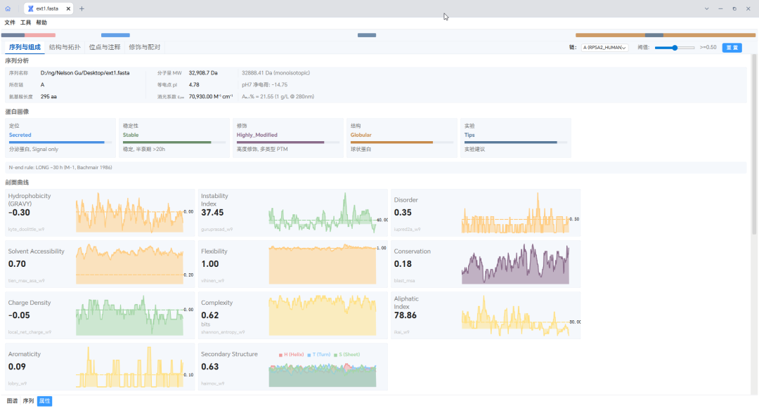
Figure 1 | D2PLab protein sequence analysis interface: from basic physicochemical properties to advanced structure prediction, visualizing molecular features.
ROUTE · CONTENTS
01PIpI is a “zero-charge” boundary line
02IEXTurning pI into a chromatography choice
03LIMITTheoretical pI is not measured pI
04PARAMSMore than pI to check before purification
05CHECKPre-purification parameter quick checklist
06EDAD2PLab: the “EDA” of the biopharmaceutical industry
The definition of the isoelectric point takes one sentence: the pH at which a protein's net charge is zero. What is easy to misread here is the word “net” — it does not mean that all dissociable groups are uncharged, but that the sum of positive charges exactly equals the sum of negative charges. Acidic residues, basic residues, and the N- and C-termini all take part in this addition.
From this definition comes a boundary line that runs through purification decisions:
Three corollaries deserve separate mention.
First, being at the pI does not mean no interaction with the medium at all. Although the net charge is zero, the charge distribution on the protein surface is uneven, and locally charged regions may still interact electrostatically with the medium; the microenvironmental pH inside the pores of the chromatography matrix may also differ from the bulk mobile phase pH. Such binding is often unstable and poorly reproducible and should not be used as a basis for method development.
Second, zero net charge means the electrostatic repulsion between molecules is at its minimum. Protein molecules can then approach one another more closely, solubility usually reaches its lowest near the pI, and the tendency to aggregate and precipitate is highest. This is exactly the principle of isoelectric precipitation, and the reason the loading pH should not sit right at the pI.
Third, pI and “net charge at pH 7” are two different parameters answering different questions. pI gives the pH at which the zero-charge point falls; net charge at pH 7 gives the actual charge state under near-physiological conditions and in conventional lysis buffers. A protein with a pI of 9.2 still carries a small positive charge at pH 7, because pH 7 lies below its pI.
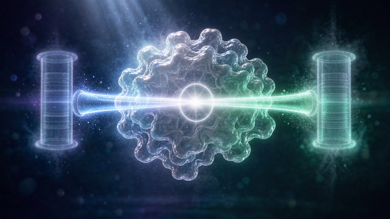
Figure 2 | Net charge reversal with pH and ion exchanger selection.
FIG 1 Net charge–pH relationship. The vertical axis is net charge and the horizontal axis is pH; the intersection of the curve with the zero line is the pI, with the right side of the intersection corresponding to anion exchange and the left side to cation exchange.
With the pI in hand, the next step is to translate it into specific media and buffer conditions. The commonly used empirical rule is:
This rule is widely cited, but if its applicable boundaries are not stated at the same time, the first loading pH a reader sets may be wrong.
Boundary one: it is a heuristic, not a law. The literature clearly points out that a considerable number of proteins do not follow this 0.5–1 unit empirical rule, mainly because the surface charge distribution of proteins is irregular; what really determines retention behavior is the spatial distribution of surface charge patches, not a scalar-form pI.
Boundary two: proteins with a pI between 6 and 8 belong to a high-risk zone. The titration curves of such proteins have a broad, flat “near-zero-charge region” around the pI, called the isoelectric region (IER) in the literature, which can span several pH units. As a result, the actual dissociation pH of proteins with a pI in 6–8 on an anion-exchange column is often significantly higher than their pI; only for proteins with a pI below 6 or above 8 is the elution pH relatively close to the pI.
A frequently cited example is Candida antarctica lipase B: only when the loading pH was brought to about 3 units below its pI was it first successfully bound to a cation-exchange medium.
Boundary three: the choice of strong versus weak media should also follow the gap between pI and pH. When the loading pH is only slightly below the pI, the protein carries only a weak positive charge, and only a strong cation exchanger (SP / S) can capture it; when the loading pH is significantly below the pI and the positive charge is sufficient, both weak and strong media can bind, and the weak cation (CM) is usually tried first. The anion side works the same way in reverse: a buffer pH slightly above the pI uses strong anion Q, and a pH significantly above the pI can use weak anion DEAE.
Boundary four: there is also a constraint that must be checked before pH — the protein's own stable pH range. The choice of medium and pH must give way to the window for maintaining enzyme activity and conformational stability: in many cases the theoretically fastest column falls exactly in the pH range that the target protein cannot tolerate.
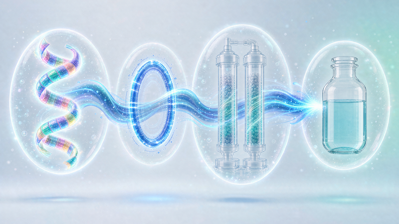
Figure 3 | The decision chain from pI to medium and buffer pH.
FIG 2 The decision chain runs from top to bottom: pI → the difference interval between loading pH and pI → strong/weak medium → re-check against the protein's own stable pH window.
Sequence-based pI prediction is essentially summing the N-terminus, C-terminus and dissociable groups such as Asp / Glu / His / Cys / Tyr / Lys / Arg according to the Henderson-Hasselbalch equation, and then numerically solving for the pH at which the net charge is zero. Treating it as a measured value is the most common failure point in this quick-reference step. The error comes mainly from three places.
The pKa sets themselves differ. Different tools use different pKa tables (the ExPASy series uses Bjellqvist values, EMBOSS uses its own Epk.dat values, and so on), and the same sequence can yield different pI values. The IPC working system published by Kozlowski in 2016 compared more than ten published pKa sets and pointed out that no single set is optimal on all datasets, and that the choice of pKa set alone can change the predicted pI of the same sequence by about 1 pH unit or even more; according to that paper, even the best-performing method at the time had an average error of about 0.87 pH units in protein pI estimation. This means that when the prediction gives 6.7, a true focusing position anywhere between 5.8 and 7.6 is not abnormal.
Post-translational modifications are not counted. Phosphorylation, glycosylation and acetylation add, remove or change dissociable groups; cysteines that form disulfide bonds no longer contribute free sulfhydryl charge. Any sequence-based prediction tool cannot see these chemical changes.
The local microenvironment is not counted. The model assumes that each dissociable group is independent and that pKa takes the model compound value. In a truly folded protein, the degree of solvent exposure, the hydrogen bond network and electrostatic interactions between neighboring charges all shift the actual pKa. The Bjellqvist pK values themselves were derived from measurements under denaturing conditions with high urea concentrations: they are more accurate for predicting the focusing position of denatured polypeptides, and less reliable for natively folded proteins. The relevant tool documentation also notes that prediction errors are usually larger for basic proteins and low-molecular-weight proteins.
Therefore, the correct way to use theoretical pI is as an anchor for setting the starting search window, not as an endpoint. After obtaining the predicted value, narrow it to an experimentally confirmed value with pH gradient elution or small-scale static binding batch screening — that is the reusable practice.
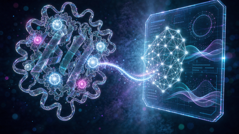
Figure 4 | Three sources of error between theoretical and measured pI.
FIG 3 Three sources of error in theoretical pI: differences in pKa sets, post-translational modifications and disulfide bonds, and local microenvironment shifts in the folded state.
A single pI cannot give a complete picture. When discussing a purification scheme, it is advisable to consider the following parameters together.
Compressing the four sections above into a checklist that can be pasted into a lab notebook.
Every purification ultimately returns to a real liquid phase. The value of the pI and this set of basic property parameters is not in making decisions for you, but in moving the starting point of the first trial-and-error a big step forward — so that limited samples and limited column runs are spent on the windows more likely to work.
Based on the world's leading cell-free protein synthesis industrialization technology, Healthcode has built the “EDA” of the biopharmaceutical industry — D2PLab molecular biology design tools — available for free download and use by research institutions and laboratories working in scientific research, drug discovery and AI protein design. D2PLab can run offline on a single machine and also supports networked computing: unified project management, AI remote large models and intelligent computing, and one-click submission for synthesis, covering the complete chain from sequence design to protein intelligent manufacturing. D2PLab has so far served more than a thousand laboratories, research institutions and AI technology companies worldwide.
Apply for a free download! Welcome to use: www.D2PLab.com